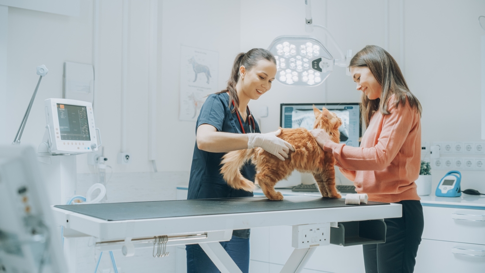
Just as in humans, pets too can develop mysterious lumps and bumps on their bodies. Often, these masses may be harmless, but sometimes they could indicate underlying health issues. As responsible pet owners, understanding the importance of timely detection and intervention is crucial. At Happy Tails Animal Hospital in Renton, WA, we’re committed to helping you navigate this often-stressful journey, ensuring the well-being of your beloved furry companion.
Why do Pets Get Lumps and Bumps? Pets can develop masses for a variety of reasons. While age is a factor and older pets tend to have more lumps, younger animals aren’t immune. These growths can arise from skin infections, insect bites, or injuries. In other cases, they could be tumors, either benign (non-cancerous) or malignant (cancerous).
Detection is Half the Battle: Regularly examining your pet at home is essential. Gently feel their body, checking for any new or changed masses. If you discover a lump, note its size, shape, consistency, and location. Is it soft or firm? Does it appear to be attached to the underlying tissue or freely movable? While not all lumps are problematic, any new or changing mass warrants a veterinary evaluation.
When to Seek Professional Help: Always consult with your veterinarian if you identify a lump on your pet, especially if:
- The mass is growing rapidly.
- It appears ulcerated or is oozing.
- Your pet seems bothered by it, often licking or scratching.
- It’s hard, immovable, and attached to deeper tissues.
At Happy Tails Animal Hospital, Dr. Arshdeep Mann and the team employ various diagnostic tools, including fine-needle aspirates and biopsies, to determine the nature of the lump.
The Process of Mass Removal: If a lump is determined to be problematic or has potential future risks, removal might be advised. Here’s a brief overview:
- Pre-Surgical Assessment: Before the procedure, your pet might undergo blood tests and imaging to assess overall health and the mass’s extent.
- Anesthesia: Since mass removals require precision and can be painful, general anesthesia is used to ensure your pet’s comfort.
- Surgery: The vet will make an incision around the mass, ensuring adequate margins to completely remove it. If it’s suspected to be cancerous, larger margins or additional tissue may be taken to ensure complete excision.
- Recovery: Post-surgery, it’s essential to monitor the incision site for any signs of infection or complications. Restrict your pet’s activity and follow any post-op care guidelines.
Post-Removal Analysis: Understanding the Significance
After the surgical removal of a mass, it’s of paramount importance to understand the nature of the growth. This isn’t just a matter of curiosity, but a vital step that directs future medical interventions and determines the best care path for your pet.
How is the Analysis Conducted? The removed tissue is preserved and sent to a specialized veterinary pathology lab. Here, the mass undergoes a process known as histopathology. In this procedure:
- Fixation: The tissue is preserved, usually using a substance like formalin, to maintain its structure.
- Sectioning: Using a microtome, extremely thin slices of the tissue are cut. This ensures that the internal structure and cells can be observed under a microscope.
- Staining: The tissue slices are treated with specific dyes that highlight different cellular structures, making it easier to identify cell types and abnormalities.
- Microscopic Examination: A veterinary pathologist examines the stained sections under a microscope. They look for the presence of cancer cells, the type of cells involved, how rapidly they’re dividing, and if they’ve invaded surrounding tissues.
What Do the Results Mean? The pathology report can offer various insights:
- Type of Tumor: Whether it’s benign or malignant, and specifics like whether it’s a carcinoma, sarcoma, etc.
- Grade of the Tumor: If malignant, the grade indicates how aggressive the tumor is likely to be. A higher grade usually implies a more aggressive growth that’s likely to spread.
- Margins: The report might comment on the surgical margins – whether any cancer cells are seen at the edge of the removed tissue. Clean margins mean that the entire tumor was likely excised, while positive margins suggest some cells might remain in the body.
Navigating the Journey Together
Your pet’s health is paramount. While the discovery of a lump can be alarming, prompt attention and intervention can make all the difference. Whether it’s a benign growth or something more serious, rest assured that with early detection and the right care, many pets continue to live happy, healthy lives post-mass removal.
If you’ve detected an unfamiliar lump on your pet or have concerns about their health, don’t delay. Reach out to Dr. Arshdeep Mann at Happy Tails Animal Hospital in Renton, WA, today. Dial (425) 254-2779 and take the first step towards ensuring your pet’s continued well-being.
Sources
- American Veterinary Medical Association. “Tumors in Pets.”
- Veterinary Cancer Society. “Lumps and Bumps in Pets: What You Need to Know.”
- Journal of the American Animal Hospital Association. “Canine and Feline Skin Tumors: Diagnosis and Treatment.”


